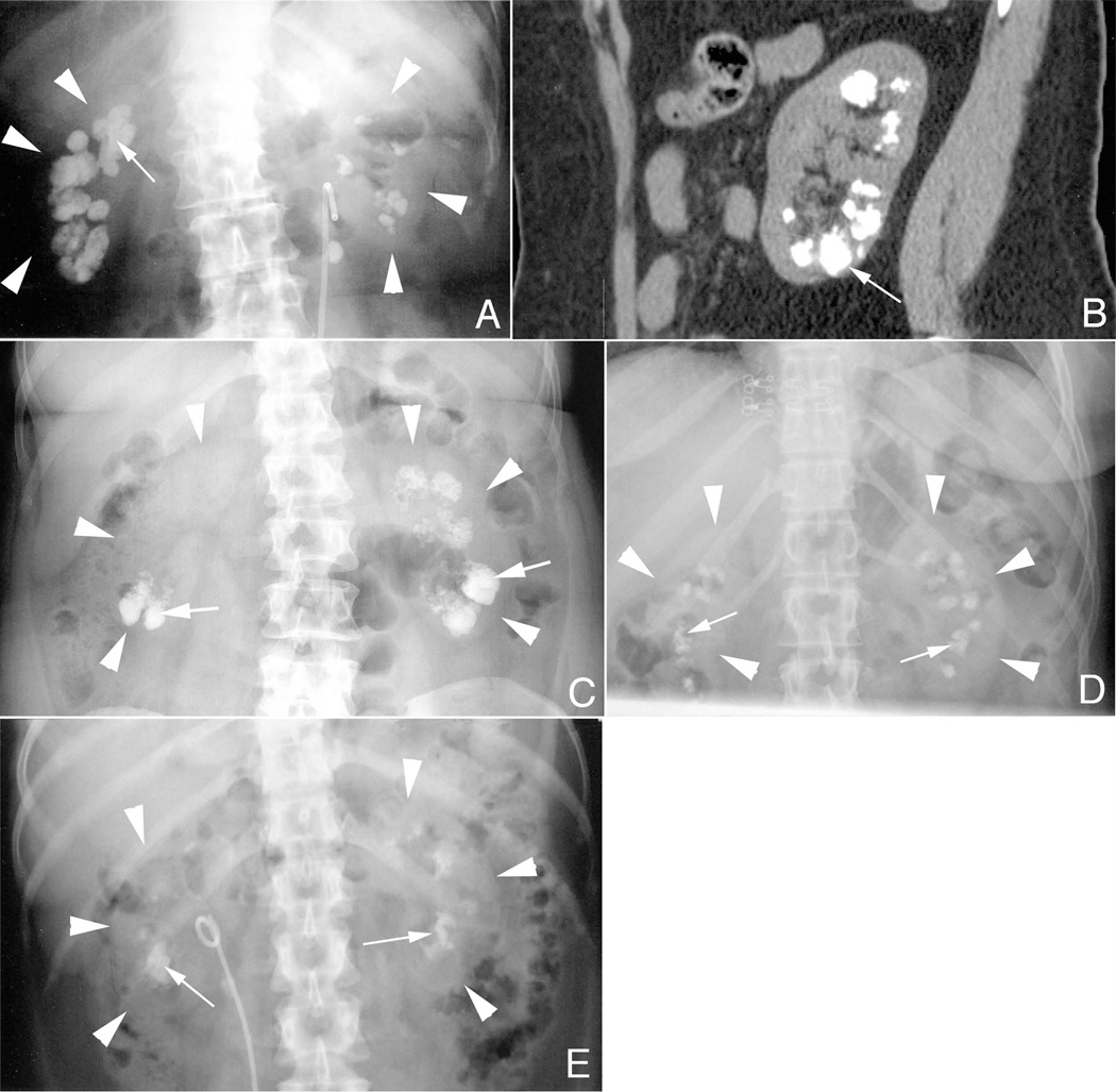
Figure 1. Radiological findings in MSK stone formers.
Panel a is a KUB from MSK patient 8 showing bilateral renal calcifications (arrow). The renal capsules of both the right and left kidneys are outlined by white arrowheads in panels a–d. By CT (panel b) some deposits (arrow) are seen extending from the cortico-medullary junction to the renal capsule. This pattern of deposits (arrows) is also seen in patients with primary hyperparathyroidism (panel c), idiopathic calcium phosphate stone disease (panel d), and distal renal tubular acidosis (panel e).

