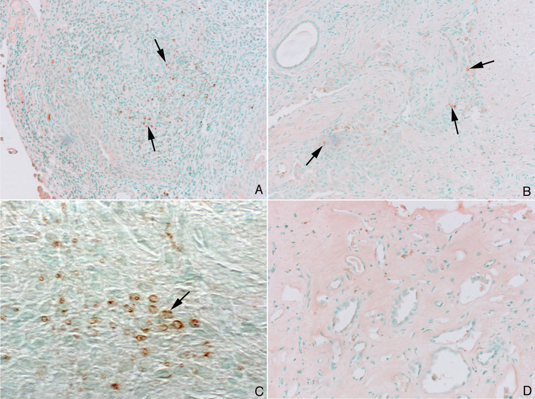
Figure 7. Immuno staining pattern for Osterix in MSK and ICSF papillary biopsies.
Like the staining pattern seen for Runx2, some nuclei of the interstitial cells in papillary section from MSK patients stained positively for osterix (panels a–c, arrows). No staining was seen in the papillary sections from ICSF patients (panel d).

