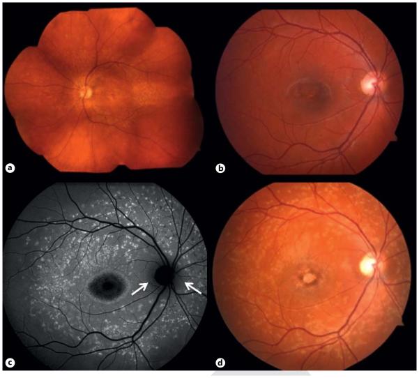
Fig. 1.
STGD. a Wide-view fundus photograph of an STGD patient. b, d Disease progression in a second STGD patient. b Fundus photograph demonstrating pigmentary changes and RPE atrophy of the fovea. FAF (c) and fundus photograph (d) after a 3-year interval showing widespread accumulation of lipofuscin and increased areas of RPE atrophy in a classic Bull’s eye shape, but with peripapillary sparing (arrows).

