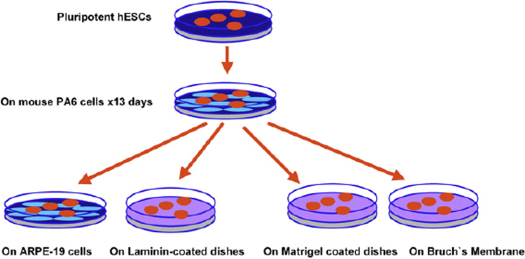
Fig. 1.
Schematic of retinal stem cell differentiation. Pluripotential hESC were grown in tissue culture dishes, harvested and seeded onto confluent monolayers of mouse PA6 stromal cells for 13 days to induce differentiation into neural progenitors. These differentiated stem cells were then harvested and cultured onto ARPE19 cell lines or laminin-coated dishes to induce photoreceptor markers; or onto human Bruch’s membrane or Matrigel to induce expression of RPE markers.

