
Figure 4.
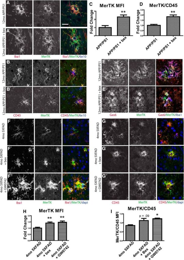
MerTK is expressed by microglia and macrophages in AD model mice and is increased by NR agonist treatment. APP/PS1Δe9 mice (12mo) were treated with vehicle (A, B, E) or the RXR agonist bexarotene (A′, B′, E′) for 7 d by oral gavage. Sections were stained for MerTK (green), Iba1 (red), and amyloid (6e10, blue) (A). MerTK mean fluorescence intensity was quantified (C). MerTK (green) colocalizes with CD45 (red), and the ratio of MerTK/CD45 mean fluorescence intensity was quantified (B, D). MerTK (green) colocalizes with Gas6 (red) and Iba1 (blue) (E). 5XFAD mice (4mo) were treated with vehicle (F, G), bexarotene (F′, G′), or the PPARδ agonist GW0742 (F″, G″) for 14 d. Sections were stained for MerTK (green) and Iba1 (red), and MerTK mean fluorescence intensity was quantified (F, H). MerTK (green) colocalizes with CD45 (red), and the ratio of MerTK/CD45 mean fluorescence intensity was quantified (G, I). Scale bar, 20 μm. Four to six animals were analyzed per group. *p < 0.05. **p < 0.01.