
Figure 1.
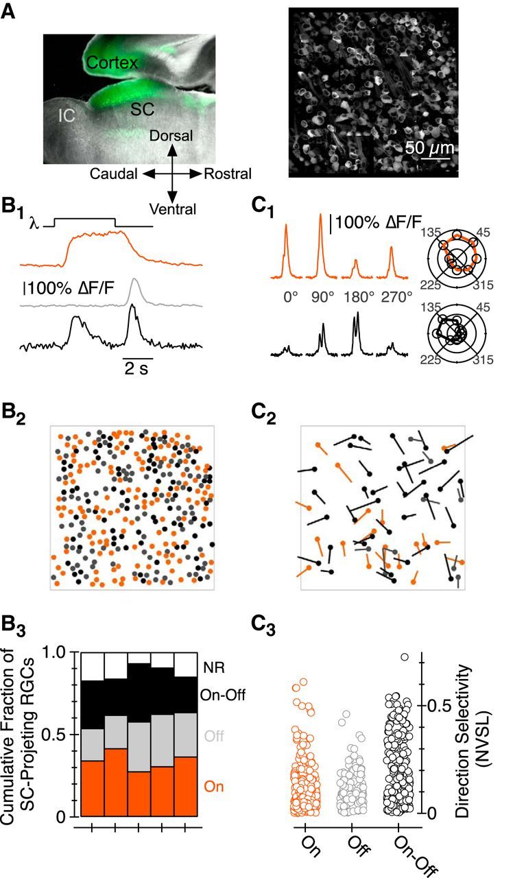
Characterization of SC-projecting RGC responses to light stimuli via GCaMP6f. A, Left, Sagittal section of a mouse brain in which AAV_hSyn_GCaMP6f was injected in the SC. Fluorescence in areas rostral to the SC originates from the axons of GCaMP-expressing SC neurons that project to pretectal nuclei; we see no fluorescent cell bodies in the pretectal areas. Right, Montage image of a subset of the RGCs labeled following injection of an AAV coding for GCaMP6f into the SC. B1, ΔF/F as a function of time in three ROIs. B2, Location of On (orange), Off (gray), and On-Off (black) SC-projecting RGCs from retina shown in A. B3, Fraction of On, Off, On-Off, and NR SC-projecting RGCs in each of five Ca2+-imaging experiments. C1, ΔF/F as a function of time for stimuli moving in one of four directions; the top/bottom data are from an On/On-Off RGC, respectively. Polar plot representation of peak ΔF/F as a function of the direction of object motion for each RGC is shown on the right. C2, Location, and preferred direction, of DS RGCs in the piece of retina shown in A; the length of each vector corresponds to the NVSL. C3, NVSL for GCaMP6f-mediated responses elicited by eight different directions of motion in each On, Off, and On-Off RGC. IC, inferior colliculus.