
Figure 1.
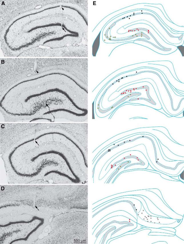
Coronal Nissl-stained sections were used to identify tetrode tracks. A–D, Arrows indicate tetrode tip locations in the dentate gyrus (A), CA3 (B), CA1 (C), and subiculum (D). A, B, Arrowheads indicate other tetrode tracks. E, Sites of recorded interneurons in the dentate gyrus (red markers), CA3 (yellow), CA1 (black), and subiculum (white) at rostral (top) to caudal (bottom) levels. Hippocampal schematic diagrams are from Paxinos and Watson (2009).