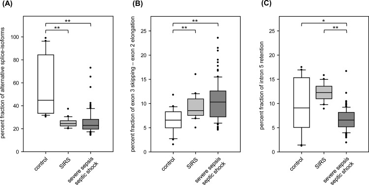
Fig 2. Characteristics of SMPD1 alternative splicing in sepsis.
Box Plots of SMPD1 splice-isoform percent fractions in control individuals (n = 20, white boxes), patients with SIRS (n = 20, light grey boxes) und severe sepsis (n = 94, dark grey boxes) for (A) the percent fraction of all alternative splice-isoforms among exons 4–6, (B) skipping of exon 3 and usage of an alternative splice donor at exon 2 (+40 nt) detected in splice-isoform ASM-2, ASM-11, ASM-24 and (C) retention of intron 5 detected in splice-isoform ASM-8, ASM-9 and ASM-11. Statistical significance (Wilcoxon rank sum test with continuity correction): * p<0.05; ** p<0.001.

