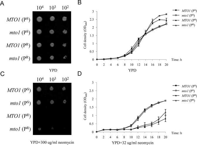
Fig 2. Phenotypes of different yeast strains.
(A) Spot assay of four yeast strains. The assay was performed by spotting decreasing concentrations of yeast cells (104,103, and 102) on a 2% glucose medium (YPD). (B) Growth curves of yeast strains in YPD for 20 hours. (C) Spot assay of four yeast strains on YPD medium containing 300μg/mL neomycin. (D) Growth curves of yeast strains on YPD medium containing 32μg/mL neomycin.

