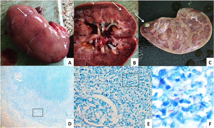
Fig 1. Macro-and microscopic images of TB in the kidney.
A. Nodules on outer surface of the right kidney and B. in the kidney parenchyma C. Tuberculoma in the kidney parenchyma D. Ziehl-Neelsen stain of a granuloma in the renal parenchyma showing glomeruli and tubules (x5) E. Microscopic zoom of indicated area with a glomerulus and multiple acid fast bacilli (x40) F. Microscopic zoom of indicated area with multiple acid fast bacilli (x100).

