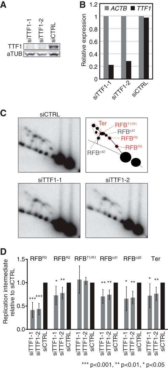
FIG 3.

TTF-1 functions in replication fork arrest at RFBs in human rDNA. (A) Levels of cellular TTF-1 as examined by Western blotting 96 h after transfection of HeLa cells with siTTF1-1, siTTF1-2, and siCTRL. (B) TTF-1 mRNA expression as examined by reverse transcription (RT)-PCR 72 h after siRNA transfection. Values were normalized to the expression level of ACTB. (C) Replication intermediates in TTF-1-depleted cells were analyzed by 2-D gel analyses 96 h after siRNA transfection. Southern blots of AflII-digested DNA were hybridized to the 28S probe. Top right, diagram showing the typical pattern of replication intermediates generated at various RFB positions (RFBT1/R1, RFBR2, and RFBR3) during head-on directional replication. RFBcd1 and RFBcd2 represent Y fork accumulation during codirectional replication at RFBR1 and RFBR3, respectively. (D) Quantification of Y forks arrested at indicated RFBs and of replication termination (Ter) detected as shown in panel C relative to the results for corresponding replication intermediates from siCTRL cells. The mean results and standard deviations (SD) from ≥6 independent experiments are shown. The P values were calculated using a one-sample t test with a hypothetical mean of 1.
