FIG 1.

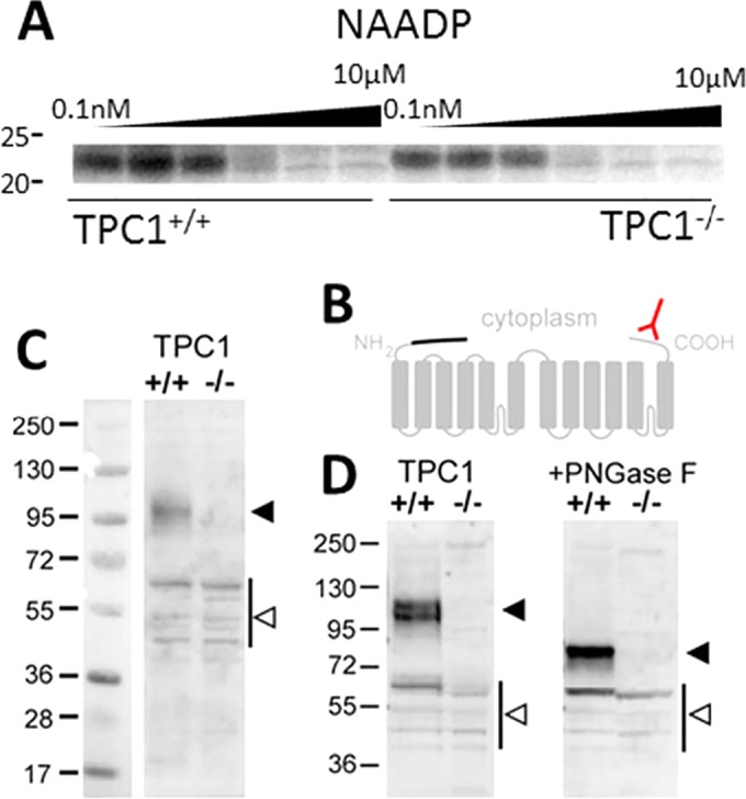
TPC1 protein is absent in transgenic TPC1 mice. (A) Photolabeling data using 5N3-[32P]NAADP showing specific photolabeling of an ∼23-kDa candidate NAADP binding protein in mouse pancreatic samples from a matched littermate mouse (TPC1+/+ [left]) and a TPC1 knockout mouse (TPC−/− [right]; Tpcn1LEXKO-471). Further detail is available in reference 11. (B) Schematic of TPC1 membrane topology to highlight the COOH-terminal epitope (red) present in wild-type TPC1 and the putative NH2-terminal truncation of TPC1B (the missing sequence is shown with a black line). (C and D) Western blot analysis of liver samples from wild-type (+/+) and Tpcn1LEXKO-471 (–/–) mice described in reference 11 using an antibody raised to the C terminus of TPC1 (ab80961; Abcam). (D) Samples were treated with peptide-N-glycosidase F (PNGase F) (right) to remove N-linked oligosaccharides as described in reference 17. Note the absence of major immunoreactive bands (solid arrowheads) in the transgenic mice. Bands present in both samples (open arrowheads) are likely nonspecific, but we cannot rule out the possibility of severely truncated, nonfunctional TPC1 variants that escape inactivation. Numbers at the left of the gels are molecular masses (in kilodaltons).
