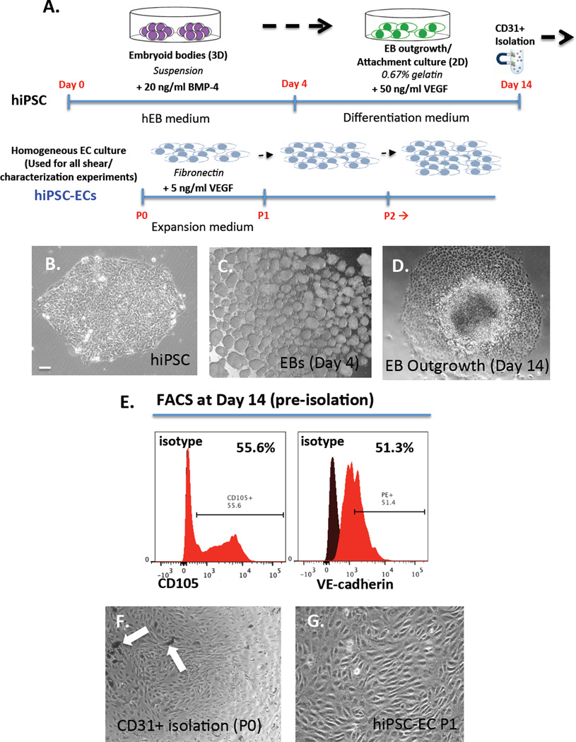
Figure 2.
(A) Schematic for directed differentiation protocol of hiPSCs to ECs in vitro in 14 days via embryoid body formation (top) and isolation/expansion of hiPSC-ECs (bottom). Phase contrast images of (B) typical hiPSC colony (C) cluster of EBs in suspension and (D) outgrowth of EBs in attachment culture at day 14 (E) FACS analysis of day 14 hiPSC-ECs (pre-isolation) showing 55.6% CD105+ cells and 51.3% VE-Cadherin+ cells (F) CD31+ magnetically isolated ECs with arrows pointing to magnetic beads and (G) isolated monolayer culture of hiPSC-ECs at passage 1 (P1). Scale bar = 10 µm.

