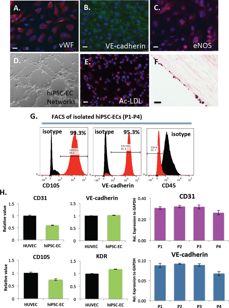
Figure 3.
Immunofluorescence analysis of EC markers in hiPSC-ECs after differentiation, isolation and expansion for 2–3 passages on fibronectin-coated plates shows expression of (A) von Willebrand Factor (vWF) (B) VE-cadherin and (C) eNOS. Functional assays demonstrate that hiPSC-ECs (D) form networks when cultured on Matrigel-coated plates for 24 hours (E) take up acetylated-LDL and (F) engraft and proliferate on decellularized human aorta slices in vitro as shown by H&E staining. Scale bars = 25 µm. (G) FACs analysis of isolated hiPSC-ECs at day 28 (P1) showing 99.3% CD105+ cells, 95.3% VE-cadherin+ cells and CD45- cells and (H) qRT-PCR analysis of EC gene expression (CD31, VE-cadherin, CD105 and KDR) in hiPSC-ECs compared to HUVECs (left) and marker expression of CD31 and VE-cadherin over multiple passages (P1–P4) in vitro (right). Values from three independent experiments from the triplicate PCR reactions for genes of interest were normalized against average GAPDH Ct values from the same cDNA sample. Fold change of GOI transcript levels between hiPSC-ECs equals 2-−ΔΔCt, where ΔCt=Ct(GOI) - Ct(GAPDH), and ΔΔCt=ΔCt(ATII) - ΔCt(ATII). (Bar indicates ± SEM and n = 3 independent experiments for qRT-PCR and flow cytometry).

