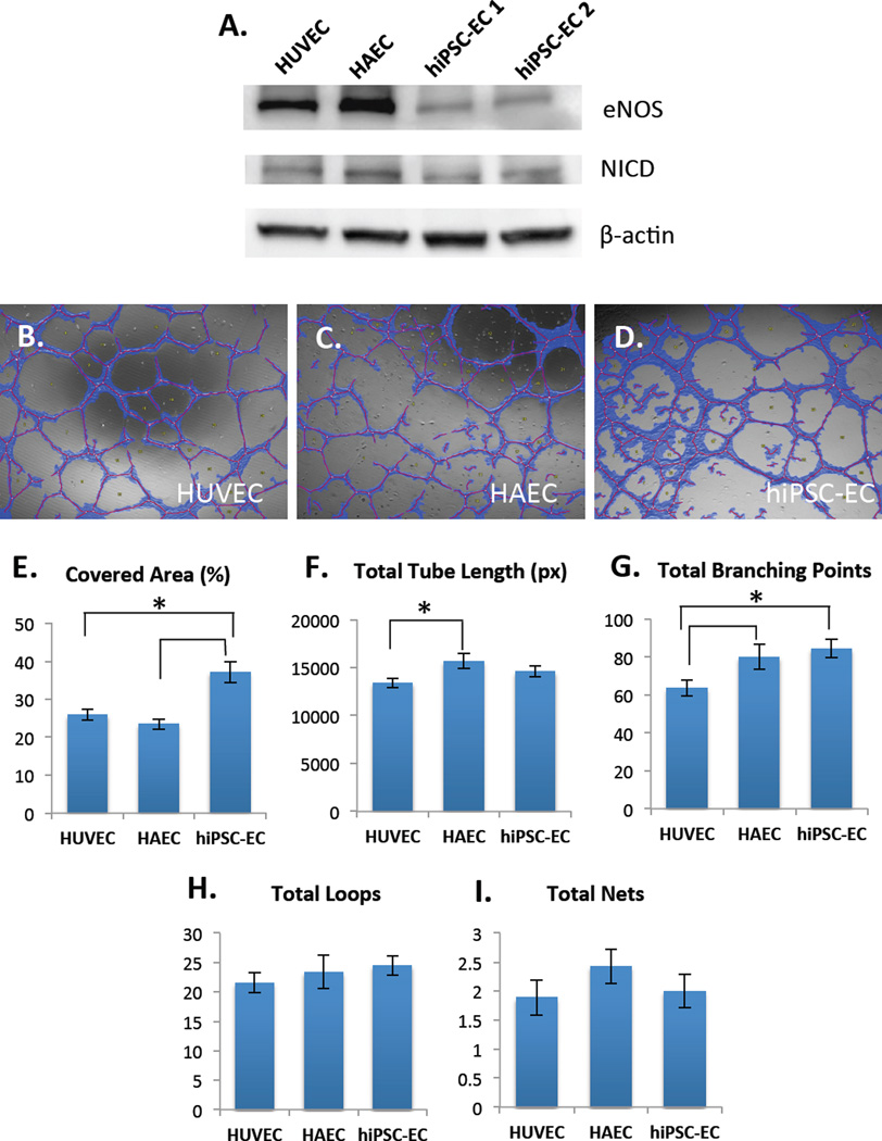
Figure 4.
(A) Western blot shows protein-level of expression of eNOS (top) and Notch1-intracellular domain (NICD) (middle) in HUVECs, HAECs and two different hiPSC-EC lines, hiPSC-EC1 and hiPSC-EC2 (under static conditions). β-actin was used as a loading control. Angiogenic properties of hiPSC-ECs (B) on Matrigel were compared to HUVECs (C) and HAECs (D) as assessed by Wimasis software. hiPSC-ECs have significantly higher covered area compared to HUVECs and HAECs (E). HAECs have higher total tube length compared to HUVECs, but not hiPSC-ECs (F). hiPSC-ECs and HAECs have significantly higher total branching points compared to HUVECs (G). There were no significant differences in total loops (H) and total nets (I). These results demonstrate certain intrinsic differences in angiogenic properties between venous, arterial and hiPSC-ECs (*p < 0.05, n=3 biological replicates, with 3 fields of view captured per sample).

