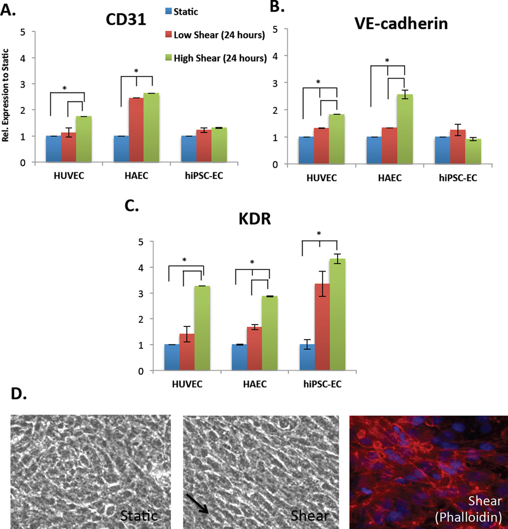
Figure 5.
The effect of shear stress (24 hours) on the gene expression, relative to static, of the mechanosensory complex comprised of (A) CD31, (B) VE-cadherin and (C) KDR in HUVECs, HAECs and hiPSC-ECs (left to right) as shown by qRT-PCR. Both low shear (red bar, 5 dyne/cm2) and high (green bar, 10 dyne/cm2) are normalized to static conditions (blue bar). All conditions are also normalized to internal house-keeping gene GAPDH. (D) Phase-contrast images of morphology of hiPSC-ECs on porous membrane in static conditions (left), alignment under shear (right, arrow pointing in direction of flow) and phalloidin showing actin stress fibers (*p < 0.05, n=3 biological replicates, n=3 technical replicates each, Student’s t-test for samples within groups).

