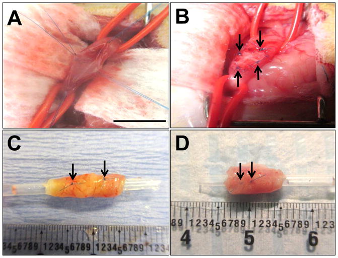
Figure 1. Rat onlay esophagoplasty model.
Photomicrographs of various surgical stages of scaffold implantation and gross morphology of regenerated tissues. [A] Esophagotomy and exposure of the esophagus lumen. [B] Anastomosis of bi-layer silk fibroin (SF) graft into the esophagus defect. [C, D] Regenerated tissues present within the original implantation sites supported by SF scaffolds [C] and small intestinal submucosa (SIS) matrices [D] at 2 m post-op. Arrows denote original marking sutures. Scale bars = 7 mm.

