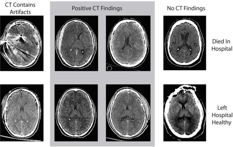
Figure 1.
Examples of varying levels of visual evidence of traumatic brain injury patients. Patients in the top row died while in the hospital and patients in the bottom row left the hospital to live at home. Patients in the first column had artifacts in their CT, patients in the middle two columns had positive CT findings, and patients in the right column had negative CT findings.

