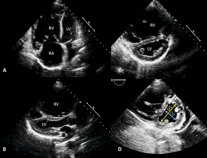
Figure 4.

Two-dimensional echocardiography of RV and LV size and ventricular interdependence. A, Apical 4-chamber view showing an enlarged RV, where the RV is larger than the LV. B, Parasternal long-axis view with an enlarged RV and bowing of the septum into the LV chamber. C, Flattening of the ventricular septum, forming a D-shaped short-axis LV appearance. D, Representation of the end-diastolic eccentricity index, which is the ratio between the LV anteroposterior dimension (D1) and LV septolateral dimension (D2). LV: left ventricle; RA: right atrium; RV: right ventricle.
