
Figure 3. Mitochondrial fusion alteration is established early during HU in gastrocnemius muscle.
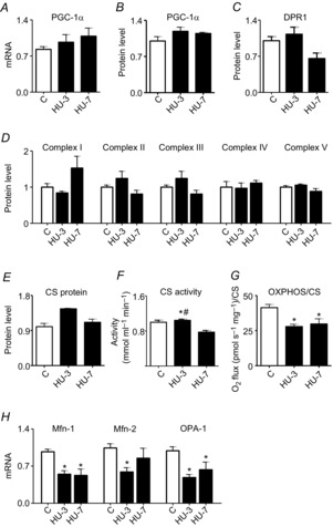
A, quantification of mRNA levels of PGC-1α by RT-PCR. B, quantification of protein levels of PGC-1α by Western blot. C, quantification of protein levels of DRP1 involved in fission machinery by Western blot. D, quantification of protein levels of mitochondrial complexes by Western blot. E, quantification of protein levels of citrate synthase by Western blot. F, determination of citrate synthase activity in skeletal muscle. G, determination of OXPHOS capacity, normalized per CS activity. H, quantification of mRNA levels of pro-fusion proteins by RT-PCR. C, control; HU-3, 3 days of hindlimb unloading; HU-7, 7 days of hindlimb unloading. *Significantly different from C, P < 0.05; #significantly different from HU-3, P < 0.05. Data are presented as means ± SEM.