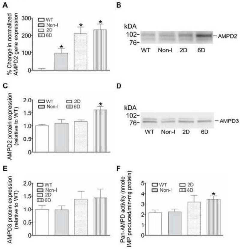
Figure 2. HIF-1α upregulates the mRNA, protein, and activity of AMP deaminase 2 (AMPD2) in mouse hearts.
Hearts were obtained from WT mice and those in which the HIF-1α-PPN transgene was suppressed (Non-I) or allowed to be expressed for 2 days (2D) or 6 days (6D). A: AMPD2 gene expression in mouse heart homogenates was examined using qPCR and normalized to reference gene transferrin. Results are expressed as the % change in gene expression relative to WT (n=6). B: Western blot showing AMPD2 protein expression in mouse heart homogenates. C: Quantification of AMPD2 protein levels in mouse heart homogenates (n=6-8). D: Western blot showing AMPD3 protein expression in mouse heart homogenates. E: Quantification of AMPD3 protein levels in mouse heart homogenates (n=6-10). F: AMPD activity assessed by the amount of IMP produced per minute per mg protein in a buffer system containing excess AMP (n=5). * P<0.05 versus WT.

