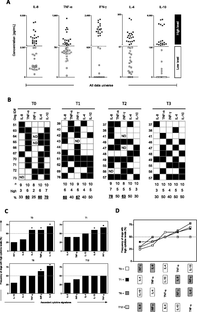
Figure 2.

Signature analysis of secreted cytokine by peripheral blood leukocytes afterin vitrostimulation withLeishmania infantum soluble antigens (SLAg). (A) Establishment of the global median cut-off edges for each cytokine (IL-8, TNF-α, IFN-γ, IL-4 and IL-10) used to segregate dogs as they present “Low” ( ) or “High” (
 ) cytokine levels. (B) Gray-scale diagrams used to compile the frequency (%) of high cytokine producers. (C) Ascendant cytokine signatures were assembled for each time after vaccination (T0, T1, T6 and T12). The frequencies of high producers were considered relevant (*) when the percentage was confined over the 50th percentile (doted lines). (D) Comparative analysis of cytokine signatures were used to identify relevant differences amongst Leishmune® vaccinated dogs at one (T1 = black rectangle), six (T6 = dark grey rectangle) and twelve (T12 = light grey rectangle) months post-vaccination compared to unvaccinated dogs (T0 = white rectangle). Gray scale rectangles were used to highlight the shift in the overall profile of pro-inflammatory and regulatory cytokines on each time after immunization. Relevant differences (shift of frequencies across the 50th percentile cut-off edge) were highlighted by gray background.
