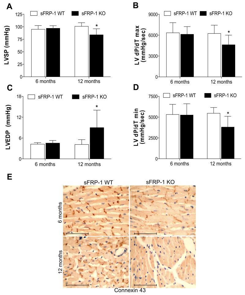
Figure 4. Deterioration of LV function in aged sFRP-1 KO mice.
Hemodynamic analysis of LV systolic (A-B) and diastolic (C-D.) function in sFRP-1 KO mice compared to their WT littermates at 6 and 12 months of age (WT/n=4); KO/n=6) representing: (A) Left Ventricular Systolic Pressure (LVSP- [mmHg]), (B) dP/dT max [mmHg/sec], (C) Left Ventricular End Diastolic Pressure (LVEDP- [mmHg]), (D) dP/dT min [mmHg]- value as below 0. Significant differences were denoted by *P < 0.05 < or **P < 0.01, or ***P < 0.001. > vs control littermate hearts. All values were expressed as mean ± SEM, (E) Immunohistochemical staining of Connexin43 in heart sections at 6 months and 12 months of age. Sections were counterstained with hematoxylin. Magnification is 40×. Scale bars represent length of 100μm.

