
Figure 2.
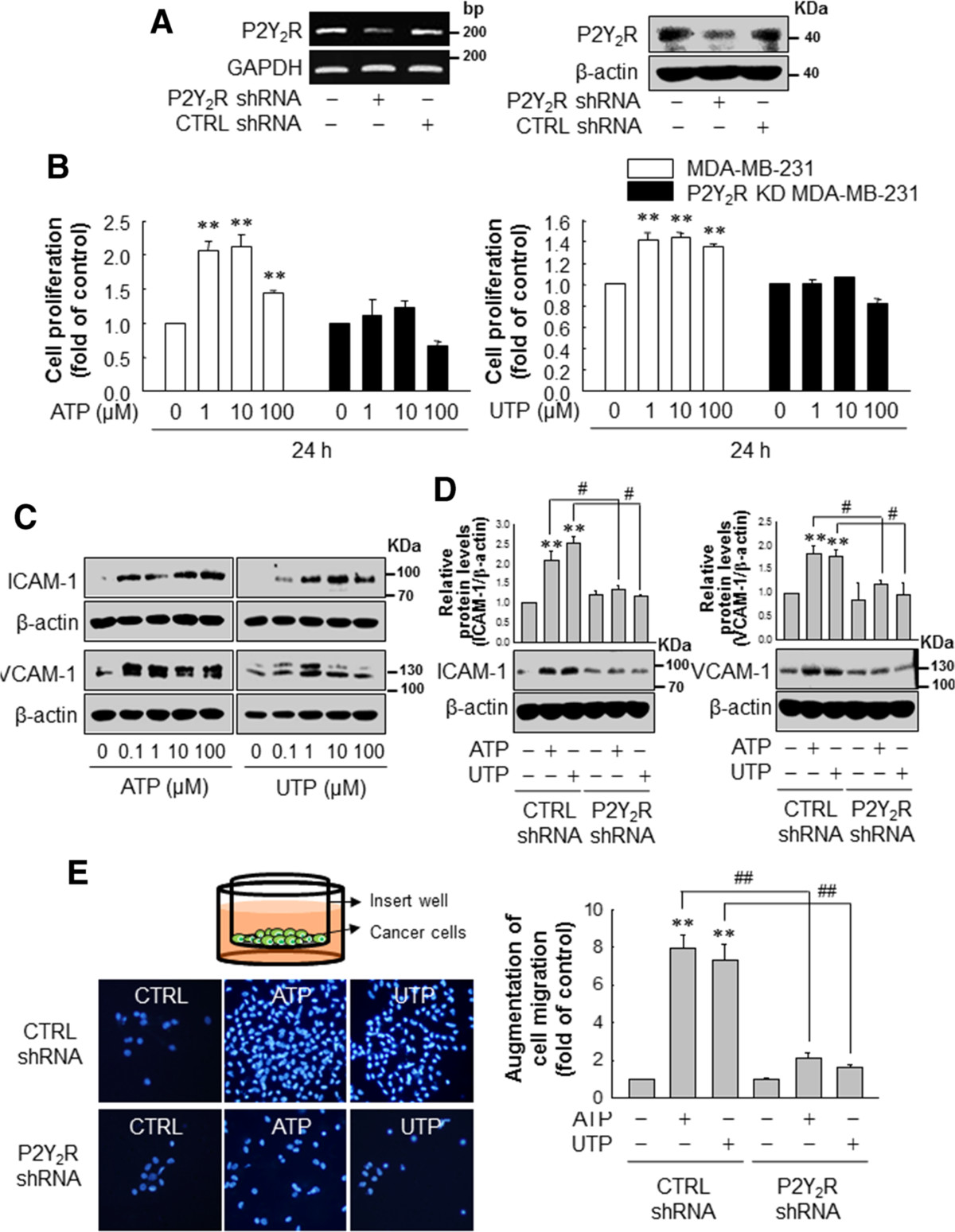
P2Y2R activation by ATP or UTP induced MDA-MB-231 cell proliferation, migration and expression of adhesion molecules. (A, B) Control- or P2Y2R-shRNA-transfected MDA-MB-231 were treated with various concentrations of ATP or UTP, as indicated. After 24 h, cell proliferation was determined by trypan blue exclusion assay. Significance compared to the control, **P <0.01. (C) MDA-MB-231 were treated with the indicated doses of ATP or UTP for 6 h. ICAM-1, VCAM-1 and β-actin expression levels were analyzed by western blotting. (D) Control- or P2Y2R-shRNA-transfected MDA-MB-231 were treated with ATP or UTP (10 μM) for 6 h, and ICAM-1 (88 to 110 KDa) and VCAM-1 (130 KDa) expression levels were determined as described previously. Significance compared to the control, **P <0.01; significance compared to ATP or UTP, #P <0.05. (E) Control- or P2Y2R-shRNA-transfected MDA-MB-231 were treated with ATP or UTP (10 μM). Six hour later, the cells were harvested, and seeded onto cell culture inserts. After 24 h, the cancer cells that had migrated across the insert well membrane were stained with DAPI, and the number of migrated cells was counted under a fluorescence microscope and quantified. Significance compared to the control, **P <0.01; significance compared to ATP or UTP, ##P <0.01.