Figure 3.
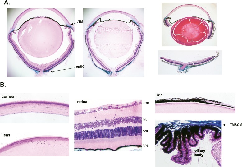
Functional recombination in Mgp-lacZ knock-in mouse line. Five-μm meridional sections from X-gal–stained MgpCre/+; R26RlacZ/+ eyes from 4- to 6-weeks old, and counterstained with hematoxylin and eosin. Sections were from three different breeding pairs of one founder. (A) Stained whole globes embedded in Technovit plastic resin (two left images) and stained bisected eyes embedded in paraffin (right image). The expression of lacZ is readily detectable in only two regions of the eye: the anterior segment TM and the posterior segment ppSC. lacZ expression is also observed in the inner layer of the midsclera. (B) Detailed meridional sections of the cornea, lens, retina, and ciliary body from the same mouse. No lacZ staining was observed in these tissues. Occasional blue staining is seen in capillaries crossing the RGC cell layer. There is a clear demarcation between the ciliary body (not stained) and the TM, heavily stained. Expression of lacZ is limited to two glaucoma-relevant tissues. Original magnifications: (A) ×40 (B) ×200. INL, inner nuclear layer; ONL, outer nuclear layer; CM, ciliary muscle.

