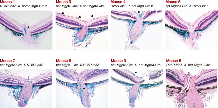
Figure 6.
Mgp Cre–mediated lacZ expression in the ONH region. Five-μm meridional sections from X-gal–stained eyes from 4- to 6-week-old and counterstained with hematoxylin and eosin. Mice 1, 3, and 4 are posterior segments of specimens shown in Figure 4. Mice 6 to 9 were from different breeding pairs. Mice 7 and 8 and 4 and 5 were littermates. Mouse 5 is the no Cre allele control (Mgp+/+; R26RlacZ/+). Eyes from mice 1, 6, 7, and 8 were stained as whole globes, with 1 and 6 embedded in Technovit plastic resin and 7 and 8 in paraffin. Mice 9 and 5 were stained as bisected specimens and embedded in paraffin. X-gal staining is very intense all across the sclera at the ppSC region indicating high expression of Mgp cre–mediated lacZ. Arrowheads: vascular smooth muscle cells from RGC vessels (mice 3 and 9) and retinal central artery (mice 1, 4, and 8) express abundant Mgp. All crosses resulted in a similar expression pattern. Original magnification: ×100.

