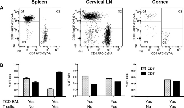
Figure 6.
CD4/CD8 ratio differs dependent on the “target tissue.” Mice received BM-TCD from B6-CD45.1 donors + T cells from B6-EGFP donors. (A) Spleens of mice that received TCD-BM + T cells displayed the characteristic GVHD phenotype, with increased number of CD8+ T cells compared to CD4+ cells, and a decreased CD4+/CD8+ ratio. Data represent analysis of cells gated on EGFP+ cells. Quadrants represent Q1, donor GFP+CD8+ (PerCP-Cy5-5A); Q3, CD4− CD8−; Q4, donor GFP+CD4+(APC-Cy7-A). Interestingly, ocular compartments such as cornea and draining cervical lymph nodes showed equal or even higher amounts of CD4+ cells compared to CD8+ T cells, creating a higher CD4/8 ratio. (B) Spleen: Data represent three to five individual spleens/group from one of two independent experiments. Cervical lymph nodes and cornea: Data represent pool of three to five mice/group from one of two independent experiments. Data are presented as fraction of total lymphocytes in each compartment.

