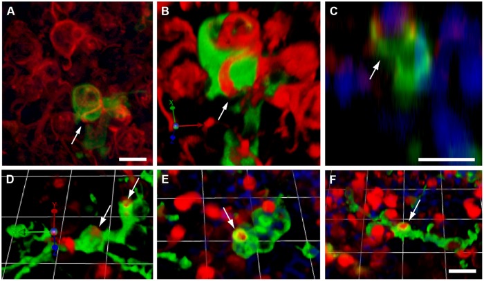
FIGURE 6.
Macrophage engulfment of hair cell debris. Confocal images of labeled hair cells and macrophages often revealed hair cells that were apparently enclosed within the processes of macrophages (arrows). (A–C) Three different renderings of the same confocal stack, showing an otoferlin-labeled hair cell (red) being engulfed by a calyx-like phagocytic process of a GFP-expressing macrophage (green). (A): Maximum intensity projection. (B) 3-D rendering of same image stack, showing the macrophage surrounding the hair cell. (C) z-plane slice from the image stack, centered on the engulfed hair cell, which is enclosed by the macrophage process (green). Cell nuclei are also visible (DAPI, blue). (D–F) Three dimensional renderings of images showing the engulfment of hair cell debris (red) by GFP-expressing macrophages (green). In all cases, phagocytic events are indicated by arrows. All scale bars represent 10 μm.

