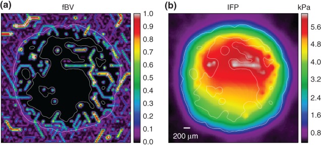
Figure 4.

Interstitial fluid flow, based on simulations of the model described in Ref 54, with a vascular network and tumor growth module as in Figure 2: (IFF) and interstitial fluid pressure (IFP) were computed for an artificially generated vascular network, incorporating a tumor vascular network in its center as a result of simulated tumor growth and vascular remodeling. Parameter settings were guided by experimental data for melanoma. The resulting network was taken as input for the IFF model. It was assumed that IFF behaves like flow through a porous medium following Darcy's law where flow velocity is proportional to the hydrostatic pressure gradient. Vessels can be sources or sinks of interstitial fluid, depending on the blood − IF pressure difference. The IFP coupling to tumor vessels was assumed to be particularly strong due to increased leakiness (permeability). A homogeneous background of lymphatics absorbs most of the flow coming from the tumor. These two plots depict a slices although the center of the tissue domain, displaying the fractional vascular volume (percentage occupied by vessels) in (a) and the IFP in (b). The location of the tumor is best inferred by the sharp drop in fBF, but it is also indicated by a thin white line. The IFP profile exhibits the expected plateaus near the center of the tumor and a steep gradient near the tumor edge. The simulation moreover predicts heterogeneities due to the particular vessel arrangement. It also shows that tumor vessels can reabsorb a significant amount of fluid if their blood pressure is much lower than neighboring vessels.
