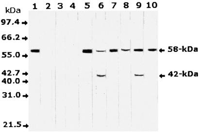
FIG. 2.
Western blot analysis of serum samples from H. pylori-infected and noninfected individuals with antisera monospecific to the 58-kDa antigen. A single band was identified at 58 kDa in serum samples from infected individuals only, in addition to a degradation product of 42 kDa in some of these samples. Lane 1, H. pylori cell lysate as a positive control; lanes 2 to 4, serum samples from three noninfected healthy individuals; lanes 5 to 10, serum samples from six individuals infected with H. pylori. Molecular mass bands are not shown but are indicated by arrows.

