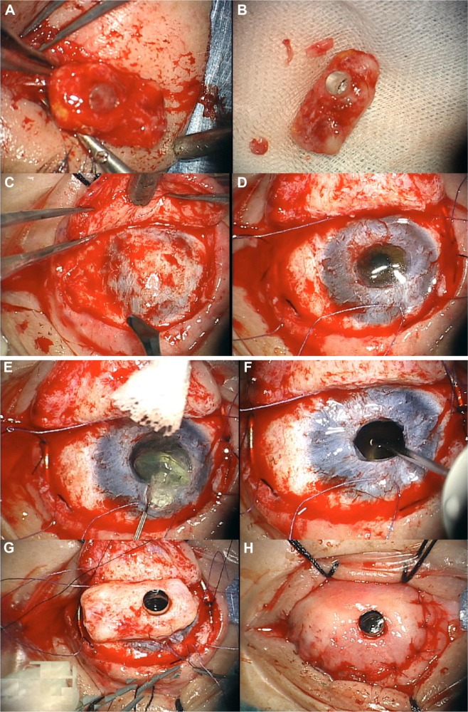
Figure 9.
OOKP stage 2 – retrieval of the lamina and implantation into the eye.
Notes: (A) Lamina is recovered from the subcutaneous pocket. (B) Connective tissue is removed from the lamina to expose the dentine side. (C) Buccal mucosa from the eye is reflected to expose the cornea. (D) Central corneal button is excised. (E) Lens extraction (IOL is also removed when present). (F) Open sky core vitrectomy is performed. (G) Lamina is implanted into the eye with wide portion of the optical stem passing through the cornea. (H) Mucosal membrane is replaced over the lamina. Through a central opening in the BMM, the optic projects beyond 1 mm.
Abbreviations: OOKP, osteo-odonto-keratoprosthesis; BMM, buccal mucous membrane; IOL, intraocular lens.

