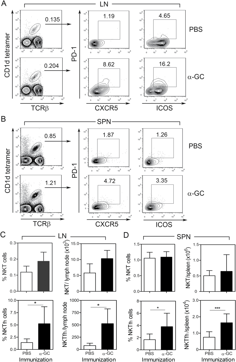
Fig. 1.
Increase in NKTfh cell numbers in vivo is stimulated by α-GC. Splenocytes and lymph node cells were harvested from B6 mice 7 days after immunization with PBS or α-GC (s.c). Cells were then stained and analyzed by flow cytometry. (A and B) Shows TCRβ+/CD1d tetramer+ NKT cells (left panels) which were further examined for expression of PD-1 and CXCR5 (middle panels) and PD-1 and ICOS (right panels). (A) Depicts lymph node cells and (B) shows splenocytes. (C) Shows mean ± SD frequency and absolute numbers of total NKT and NKTfh cells in lymph node. (D) Shows mean ± SD frequency and absolute numbers of total NKT and NKTfh cells in spleen. Data shown in A–D are each representative of two independent experiments using five mice per group. Statistically significant differences between experimental groups were determined using a two-tailed unpaired t-test (*P < 0.05, ***P < 0.001).

