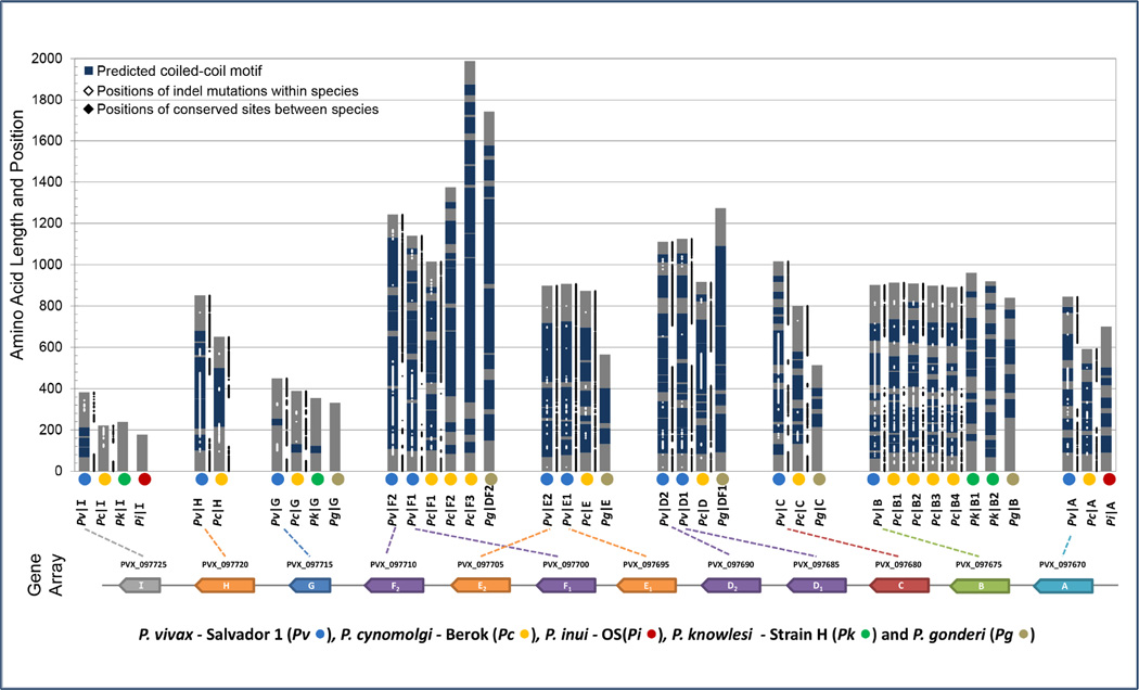
Figure 3.
Domain architecture of the Pvmsp3 family and its orthologs in related species. Vertical gray bars represent putative MSP3 proteins, with their amino acid lengths drawn to scale. The positions of coiled-coil tertiary motifs as predicted by PairCoil2 are shown in blue. Positions of sites within indels from intraspecies alignments of P. vivax and P. cynomolgi isolates are plotted onto the proteins in white along with the positions of sites conserved and alignable between P. vivax and P. cynomolgi (in black). The P. vivax Salvador-1 gene array is shown for reference along with the abbreviations used for the nonhuman primate malaria species analyzed.

