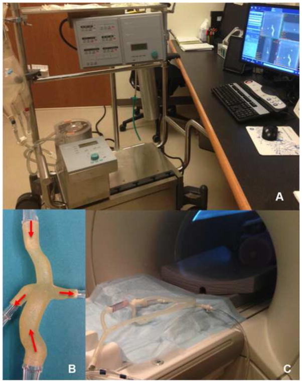
Figure 3.
Physical models were connected to a perfusion system (A), which circulated water entering through the vena cavae and exiting through the pulmonary arteries as shown for the extracardiac TCPC case (B). The physical model and tubing were placed in the scanner (C) to assess fluid flow by means of 4D Flow MRI.

