Figure 4.
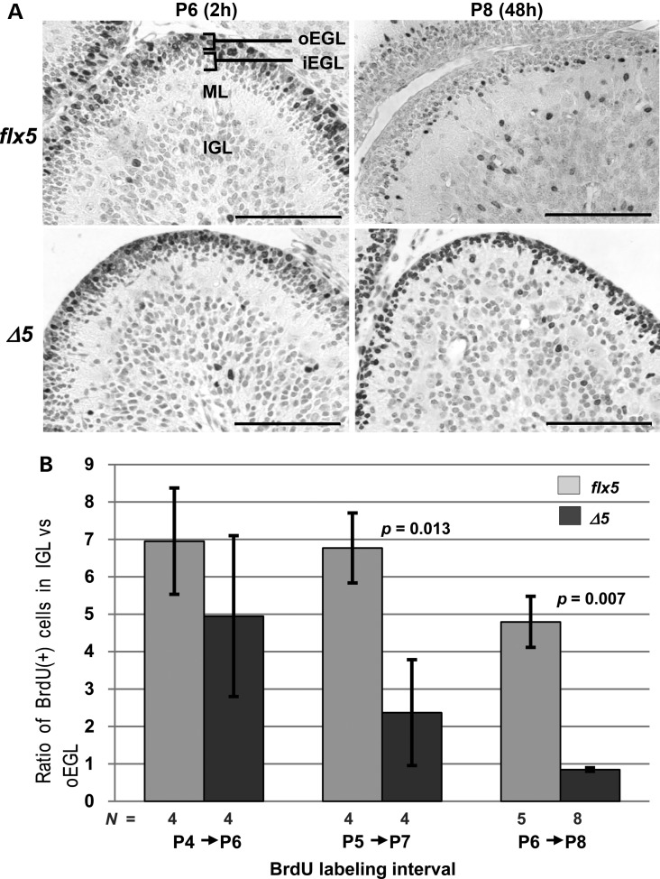
Migration of BrdU-labeled GCPs in Nsdhlflx5/Y and NsdhlΔ5/Y cerebella. (A) Distribution of BrdU-labeled cells in the EGL of lobule 3 in Nsdhlflx5/Y and NsdhlΔ5/Y cerebella at 2 and 48 h after BrdU injection on P6. The lighter staining of cells in the Nsdhlflx5/Y sample at P8 was likely due to dilution of incorporated BrdU by cell division that followed the initial labeling 48 h earlier. Note that a large majority of the labeled GCPs in the NsdhlΔ5/Y sample remained in the oEGL in the P8 sample, 48 h post-injection. Abbreviations: oEGL, outer external granule layer; iEGL, inner external granule layer; ML, molecular layer; IGL, inner granule cell layer. Scale bars: 100 μm. (B) The migration efficiency of Nsdhlflx5/Y and NsdhlΔ5/Y GCPs at different ages. Migration was quantified by injecting pups with BrdU at P4, P5 or P6 and collecting cerebella for IHC 48 h later at P6, P7 or P8, respectively. The ratio of BrdU-labeled cells in the IGL to those remaining in the oEGL of lobule 3 was calculated for each sample (see Methods).

