Figure 5.

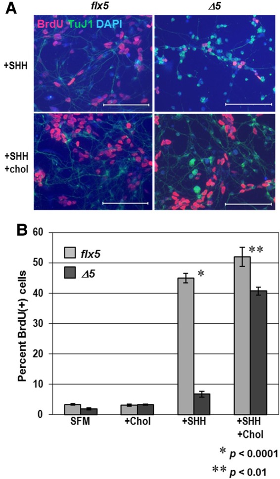
Response of cultured GCPs to SHH and exogenous cholesterol. (A) Immunofluorescent staining for BrdU (red) and neuronal marker TuJI (green), with DAPI (blue) counterstaining of Nsdhlflx5/Y and NsdhlΔ5/Y GCPs that were cultured for 48 h with either 1 μg/ml recombinant SHH alone or SHH plus 15 μg/ml cholesterol. BrdU was added to the samples 4 h before fixing. Note that there are fewer and thinner neurite extensions in the mutant NsdhlΔ5/Y cells than in the Nsdhlflx5/Y controls. Scale bars: 100 μm. (B) Four aliquots of isolated GCPs from individual Nsdhlflx5/Y and NsdhlΔ5/Y P4 cerebella were cultured for 48 h in either serum free medium (SFM) alone, SFM with 15 μg/ml cholesterol (chol), SFM with 1 μg/ml recombinant SHH, or SFM with 15 μg/ml cholesterol and 1 μg/ml SHH. BrdU was added to the cultures 4 h before fixing the cells. Cells were immunostained for BrdU and counterstained with DAPI. Values are the percentage of BrdU-labeled cells from the total (DAPI-stained) number of cells. Each bar represents the mean ± SEM of results from three independent experiments.
