Figure 1.
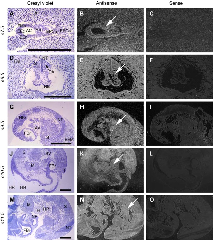
Mbtps1 is expressed ubiquitously in the post-implantation and mid-gestation mouse embryo. Left column shows embryo sections stained with Cresyl Violet; middle column shows the same sections with comparative antisense hybridization labeling seen as bright under darkfield illumination (arrows) and right column shows control sense hybridization. (A–C) Comparison of in situ hybridization with sense and antisense probes reveals a broad expression of Mbtps1 in the uterine decidua and in the embryo at E7.5 (arrow). E7.5 early embryo sagittal section seen within uterine cavity near site of implantation. Presence of Mbtps1 mRNA labeling is evident within embryo, ectoplacental cone and decidua. (D–F) E8.5 embryo coronal section. (G–I) E9.5 embryo sagittal section. (J–L) E10.5 embryo sagittal section. (M–O) E11.5 embryo sagittal section. AC, amniotic cavity; AV, atrioventricular canal; DA, dorsal aorta, primordium; De, decidua; EEc, embryonic ectoderm; EEM, extraembryonic membranes; EEn, embryonic endoderm; Em, embryo; EMe, embryonic mesenchyma; EPCa, ectoplacental cavity; EPCo, ectoplacental cone; FBr, forebrain; H, heart; HBr, hindbrain; HP, hepatic primordium; HR, hinbrain roof; M, mandible; NE, neuroepithelium; NP, nasal pit; NT, neural tube; S, somite; SV, sinus venosum; T, tail; and Tr, trophoblasts. Magnifications: (A–C) ×36; (D–F) ×24; (G–I) ×15 and (J–O) ×10. Scale bar: 1 mm.

