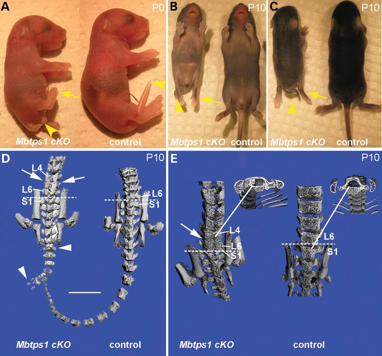
Figure 2.
Mbtps1cKO mice exhibit a severe caudal truncation and changes in vertebrae patterning. (A) At P0, Mbtps1cKO mice exhibit caudal axial truncation (arrowheads) and hypotrophy of the hind limbs (arrows). (B and C) At P10, Mbtps1cKO mice are smaller than normal littermates and the caudal axial truncation, as well as the hypotrophy of the hind limbs, are even more evident. B, ventral and C, dorsal view. (D and E) Micro-CT images of control and Mbtps1cKO mice at P10. (Each micro-CT image represents a separate mouse.). (D) Posterior comparison of Mbtps1cKO spine with that for control littermate. Arrowheads denote abnormal tail vertebrae and arrows demark abnormal bone patterning and fusion of lower lumbar vertebrae. Scale bar: 5 mm. (E) Sacral-lumbar comparison. Arrow denotes region with abnormal bone patterning and fusion of L4-L6 vertebrae in Mbtps1cKO. Solid lines identify regions from which respective cross-sections were taken. Dashed lines denote the sacral-lumbar border. S, sacral. L, lumbar.

