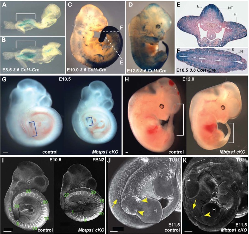
Figure 3.
The Cre recombinase expression in 3.6 Col1-Cre mice nicely correlates with the caudal phenotype of the Mbtps1cKO mice. (A–D) Whole-mount LacZ staining of 3.6 Col1-Cre;ROSA26 mice at E8.5 (A,B), E10.0 (C) and E12.5 (D) reveals expression of the Cre recombinase prominently in the posterior tail region (brackets). (E and F) LacZ stained transverse sections of E10.5 3.6 Col- Cre: ROSA26 embryos showing strong expression of the Cre recombinase in the somites (S), the neural tube (NT), the hind limb buds (H) and the ectoderm (E). Sections were taken at the axial levels denoted in 3C. (G and H) A caudal axial truncation is evident in homozygous Mbtps1cKO embryos (right) by E10.5 compared to their wild type (left) littermates (G, brackets). The caudal axial truncation becomes more prevalent by E12.0 (H, brackets). (I) At E10.5, Fibrillin2 (FBN2) antibody staining reveals a reduced number of somites in Mbtps1cKO embryos (right) compared to their littermate control (left) (27 instead of 30 in these embryos). (J and K) Whole mount antibody staining of neuron-specific class III β tubulin (TUJ1) at E11.5 in Mbtps1cKO embryo (K) and control littermate (J) reveals a general failure of neuronal projections in the Mbtps1cKO embryos (arrows). Interestingly, no neuronal projections are seen in the hind limb bud (H) and spines of Mbtps1cKO embryos (arrowheads). Scale bar: 500 μm.

