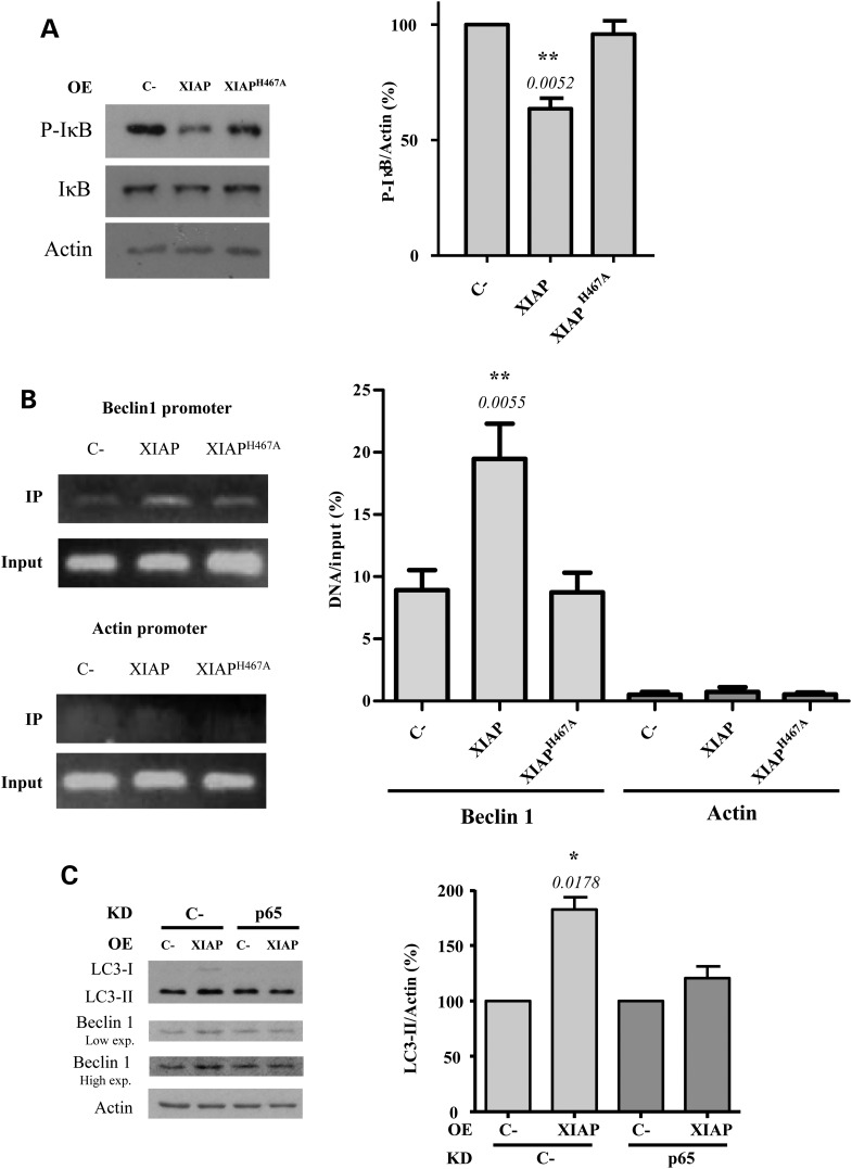
Figure 4.
p65 is involved in the activation of autophagy by XIAP. (A) HeLa cells previously transfected with empty vector (C-), wild-type XIAP or XIAPH467A expression constructs for 48 h were subjected to western blotting to detect P-IκB and IκB levels. The blots are from the same set of experiments. Densitometric measurements of phospho-IκB (P-IκB) bands were normalized to the corresponding bands of actin and are shown in the histogram on the right. (B) HeLa cells previously transfected with empty vector (C-), wild-type XIAP or XIAPH467A expression constructs for 48 h were subjected to a ChiP assay. The amount of in vivo binding of endogenous p65 to Beclin 1 and actin (as a negative control) promoters was quantified by real-time PCR. Data are representative of three independent experiments. (C) HeLa cells were transfected for 48 h with a control (C-) or p65 siRNA. Twenty-four hours later, cells were co-transfected for 48 h with an empty vector (C-) or XIAP expression constructs. Cells were treated with DMSO or 400 nm bafilomycin A1 during the last 4 h. Densitometric measurements of LC3-II bands were normalized to the corresponding actin bands and are shown in the histograms on the right. The values shown in all the histograms represent the mean ± standard deviation from at least three independent experiments performed in triplicate samples/condition. The P-values were determined using Student's t-test. See also Supplementary Material, Figure S4.

