Figure 1.
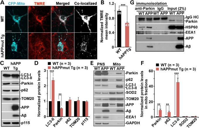
AD-linked mitochondrial stress induces mitophagy in mutant hAPP Tg neurons. (A and B) Representative images (A) and quantitative analysis (B) showing accumulation of depolarized mitochondria in mutant hAPP Tg neurons. Cortical neurons cultured from WT or mutant hAPP Tg mice were transfected with mitochondrial marker CFP-Mito, followed by loading with mitochondrial membrane potential (Δψm)-dependent dye TMRE for 30 min prior to imaging. TMRE mean intensity was normalized to WT neurons. Note that hAPP mutant neurons displayed reduced TMRE mean intensity in the soma relative to that of WT neurons. Scale bars: 10 μm. Data were quantified from a total number of neurons as indicated in the bars (B) from > 3 independent experiments. (C and D) Representative blots (C) and quantitative analysis (D) showing altered mitophagy/autophagy markers in the brains of mutant hAPP Tg mice. A total of 20 μg of brain homogenates from WT and mutant hAPP Tg mice were sequentially detected on the same membrane. Relative protein levels were normalized by Golgi marker p115 and compared with that of WT littermates. (E and F) Increased mitochondrial association of Parkin, p62/SQSTM1 and LC3-II in the brains of mutant hAPP Tg mice. Following Percoll-gradient membrane fractionation, equal amount (5 μg) of mitochondria-enriched membrane fraction (Mito) and post-nuclear supernatant (PNS) from WT and mutant hAPP Tg mice were sequentially immunoblotted with antibodies against Parkin, autophagy markers p62 and LC3, mitochondrial markers SOD2 and TOM20, early endosome marker EEA1 and cytosolic protein GAPDH, along with APP and Aβ (6E10). The purity of Mito fractions was confirmed by the relative enrichment of mitochondrial markers SOD2 and TOM20 compared with PNS fractions, and by the absence of EEA1 and GAPDH. Relative protein levels in mitochondria-enriched membrane fraction of AD mice were compared with those in WT littermates. (G) Immunoisolation assay showing association of APP and Aβ with Parkin in mutant hAPP Tg mouse brains. Parkin-associated membranous organelles were immunoisolated from light membrane fractions with anti-Parkin-coated Dyna magnetic beads, followed by sequential immunoblotting on the same membranes after stripping between each antibody application. Data in C, D, E, F and G were analyzed from three pairs of mice for each genotype and expressed as mean ± SEM with Student's t-test: ***P ≤ 0.001; **P < 0.01; *P < 0.05.

