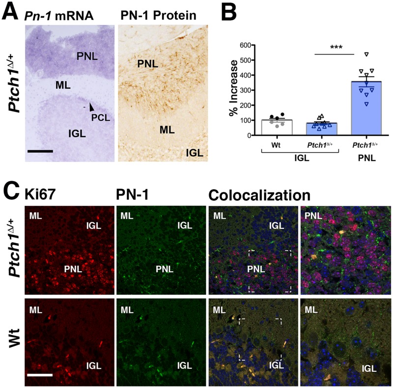
Fig 2. Serpine2/PN-1 is overexpressed in cerebellar PNLs of Ptch1 Δ/+ mice.
(A) Left panel: Serpine2/Pn-1 transcript distribution (purple staining of the RNA hybrids) in the cerebellum of a Ptch1 Δ/+ mouse 6 weeks after birth. Expression is highest in the pre-neoplastic lesions (PNL) within the cerebellum. Right panel: Serpine2/PN-1 protein levels (brown staining of the immunocomplexes) are highest in the PNLs of Ptch1 Δ/+ mice. (B) qPCR analysis of Serpine2/Pn-1 transcripts in laser-dissected tissue from wild-type (Wt) and Ptch1 Δ/+ normal IGL tissues in comparison to Ptch1 Δ/+ PNLs. Expression in the wild-type IGL was set to 100% (S3 Table), p≤0.001. (C) Co-localization of Ki67-positive proliferating cells (red fluorescence) and Serpine2/PN-1 (green fluorescence) in the cerebellum of wild-type and Ptch1 Δ/+ mice at 6 weeks (n = 3). White lines indicate the position of the enlargement shown in the right-most panel. Auto-fluorescent cells are detected equally in the red and green channel and appear yellow-orange in the overlap (Colocalization, right panels). IGL: internal granular layer; ML: molecular layer; PCL: Purkinje cell layer; PNL: pre-neoplastic lesion. Scale bars: 100μm.

