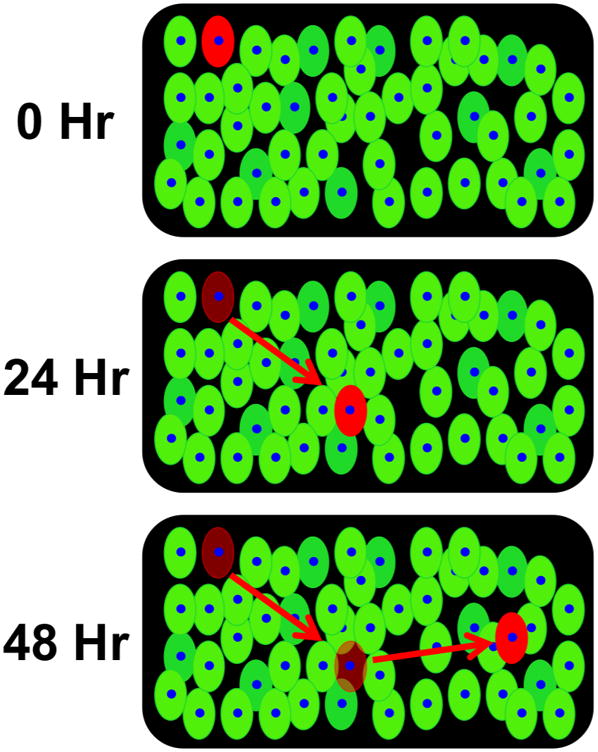
Fig. 5. Schematic diagram of cell tracking in tissue culture.

The diagram illustrates how cell tracking could work in a tissue culture if a single transfected cell is photo-converted. The photo-converted cell would appear red against cells that are green, which would allow for precise tracking of cell movement and observation of morphological changes in tissue culture.
