
Figure 1. Induction of Parkinsonism by intracranial administration of 6-OHDA.
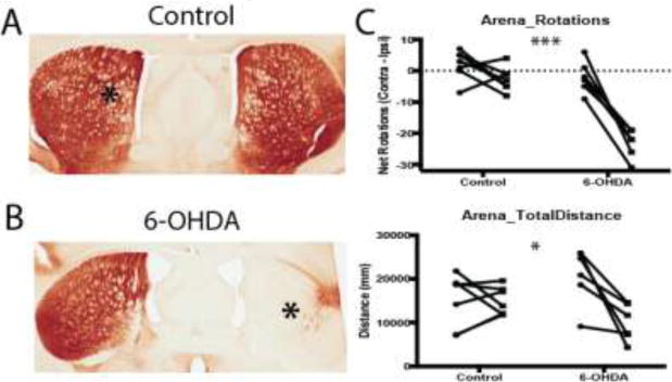
A–B. TH immunostaining in the striatum after unilateral administration (marked with an asterisk) of saline (A) or 6-OHDA (B) into the MFB, demonstrating striatal dopamine depletion on the 6-OHDA-lesioned side. This resulted in significant behavioral deficits as shown in the open-field test (C). 6-OHDA-lesioned mice spontaneously rotated in the ipsiversive direction (side of the lesion) and traveled a shorter distance in the arena as compared to control mice. For each group, behavioral measures collected before and after injection for each mouse are shown as connected data points. *, p < 0.05; ***, p < 0.001