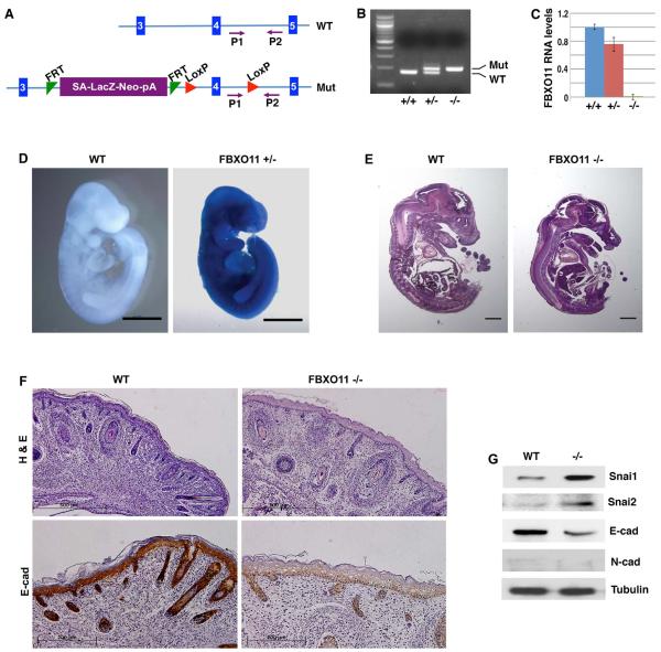
Fig. 6. Generation and characterization of FBXO11 mutant mice.
(A) Schematic representation of FBXO11 inactivation. The mutant allele (Mut) contains a “LacZ-Neo” cassette that also includes the mouse En2 splice acceptor (SA) and the SV40 polyadenylation sequences (pA) [37]. This cassette acts as an exon, which stops the transcription of FBXO11 at the pA and thus creates a null allele. Blue vertical bars represent the exons of FBXO11. The exon 4 encodes the F-box motif. FRT and LoxP are specific DNA elements for potential recombination and conditional deletion (irrelevant in the current study). Primers P1 and P2 are used for genotyping.
(B) Genotyping of mouse FBXO11 mutants. Wild type (WT), FBXO11 heterozygous (+/−) and homozygous (−/−) mutant mice were genotyped by PCR with the P1 and P2 primers (WT: 179 bp; Mut: 204 bp).
(C) Validation of FBXO11 deficiency in the mouse FBXO11 homozygous mutants. Mouse E10.5 embryos with indicated genotypes were analyzed for FBXO11 RNA expression by quantitative RT-PCR (with primers corresponding to the exons 3 and 4). Expression of FBXO11 in the homozygous mutant was undetectable (less than 0.1% of the WT).
(D) Whole mount β-Galactosidase staining of wild type (WT) and FBXO11 heterozygous (het) E10 mouse embryos. FBXO11 mutant allele carries a knock-in LacZ reporter. Scale bar: 1mm.
(E) Sagittal sections of WT and FBXO11 homozygous mutant (KO) E15.5 mouse embryos (H&E, scale bar: 1mm).
(F) FBXO11-deficient mouse epidermis exhibits reduced hair follicles, thickened epidermis and diminished E-cadherin expression. Sagittal sections of newborn wild type (WT) and FBXO11 −/− littermate were stained with H&E (scale bar: 500μm) or immunohistochemically with an anti-E-cadherin antibody (scale bar: 200 μm).
(G) Elevated Snai1 protein levels in FBXO11 mutants. Protein extracts were prepared from the back skin of neonatal WT and FBXO11 KO embryos, and immunoblotted with indicated antibodies.

