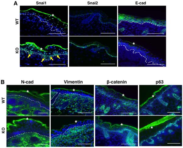
Fig. 7. Molecular characterization of epidermis from FBXO11-deficient mouse embryos.
Cryosections of FBXO11 mutant and control littermate embryos at E18.5 were stained for immunofluorescence analysis with antibodies specific for Snai1, Snai2, and E-cadherin (A) as well as N-cadherin, Vimentin, β-catenin, and p63 (B). Dashed lines demarcate the basement membrane that separates the epidermis from dermis. Arrows indicate basal cells with increased Snai1 expression. The asterisks denote autofluorescence of the cornified layer. Scale bar: 100μm.

