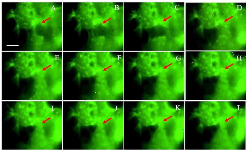
Fig 3. The fluorescence observation for the live Z172 cells imaging by time-lapse recording.
The fluorescent observation of the progress of Z172 cells which was transfected pAcGFP1-Actin after co-cultured with T. vaginalis in 1:10. Panel A to L were the captured images once every 30 minutes. The arrows indicate the position of the cell gap.

