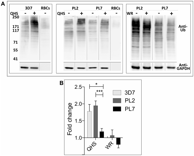
Fig 4. Ubiquitination of P. falciparum proteins following ART treatment.
Uninfected RBCs or trophozoite-infected RBCs (24–44 h p.i.) (3% hematocrit) of 3D7, PL2, and PL7 strains were incubated with 1 μM QHS or 20 nM WR99210 for 90 min at 37°C. Cell extracts were subjected to SDS-PAGE and western blotting and probed with anti-ubiquitin IgG with ECL detection, then stripped and re-probed with anti-PfGAPDH. (A) Representative western blots. (B) Densitometric analysis of the anti-ubiquitin signal for at least nine QHS and three WR99210 experiments. Significance was determined using a Student’s t test. * p <0.05; *** p <0.005.

