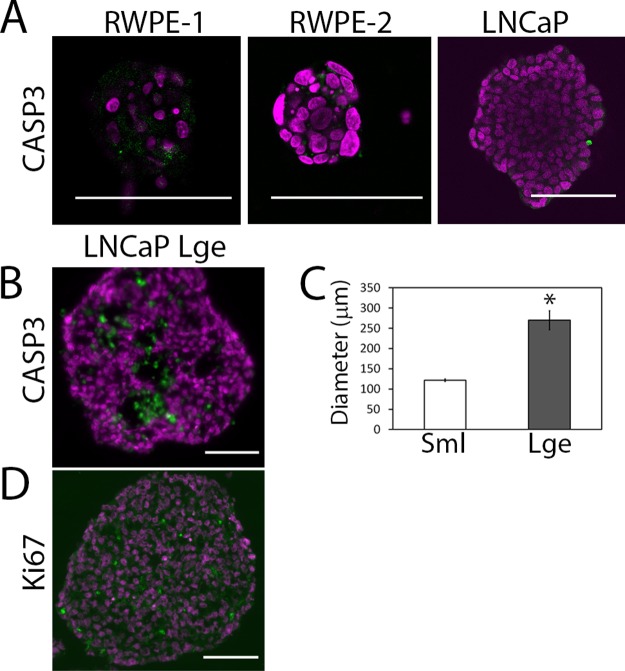
Fig 5. Apoptotic core can be controlled for using differently sized microwells.
(A) Cleaved caspase-3 (CASP3; green), an apoptosis marker, was used to stain sections of RWPE-1, RWPE-2 and LNCaP microaggregates. (B) Due to the absence of cleaved caspase-3 in the small LNCaP aggregates, a large microwell (800 μm ×800 μm ×800 μm) was used to create large LNCaP microaggregates with an apoptotic core (CASP3 Lge). (C) The diameter of the LNCaP microaggregates (Sml) was compared with LNCaP microaggregates grown in the large microwells (Lge). A minimum of 50 aggregates were measured per condition. (D) Ki67 (green) was also used to stain proliferating cells within the large LNCaP aggregate. Nuclei were stained with DAPI (magenta); scale bar is 100 μm.

