
Figure 2.
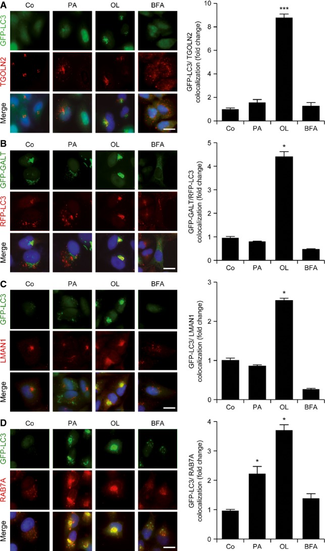
Subcellular localization of autophagic markers during fatty acid-induced autophagy
- A–D Colocalization of GFP-LC3+ dots with the Golgi apparatus in cells exposed to oleate. GFP-LC3- or GFP-GALT/RFP-LC3-expressing U2OS cells were maintained in control conditions (Co) or treated with 500 μM palmitate (PA), 500 μM oleate (OL) or 10 μg/ml brefeldin A (BFA) for 6 h. Thereafter, cells were processed to determine the colocalization between GFP-LC3+ or RFP-LC3+ dots and TGOLN2 (A), GALT-GFP (B), LMAN1 (C) and RAB7A (D). Data are normalized means ± SEM of at least three independent experiments (*P < 0.05, ***P < 0.001 versus untreated cells). Scale bars, 10 μm.